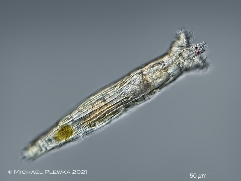
| 1. Rotaria tridens; characteristic is the protruded rostrum while swimming and feeding, which is in contrast to Rotaria rotatoria. (1) |
| |
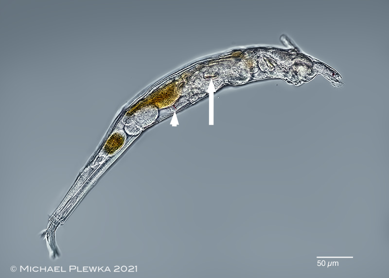 |
| 2. Rotaria tridens; lateral view of pregnant specimen with retracted corona. The daughter can be recognized by the eyespots marked by the arrowhead) and the trophi (marked by arrow). (1) |
| |
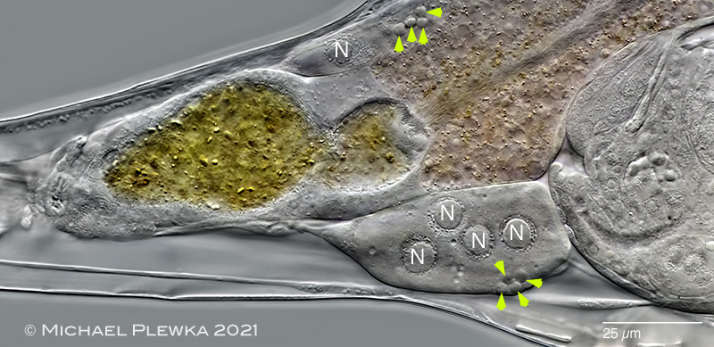 |
| 3. Rotaria tridens; the germovitellarium in the lower half of the image shows 4 nuclei (N) of the vitellarium (the smaller ones are oocytes from the ovary (green arrowheads; although only 4 oocytes are visible in this image the actual number of oocytes is ≥ 8 in this morphotype). This observation is also supported by a video that scans through the whole germovitellarium. All observed specimens from the morphotype of this sample had 4 nuclei. |
| |
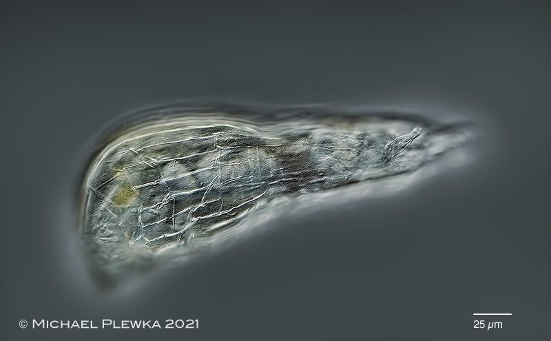 |
| 4. Rotaria tridens; creeping specimen, focus plane on the integument and dorsal antenna. |
| |
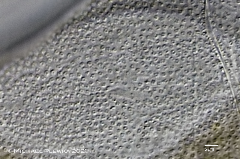 |
| 5. Rotaria tridens; granulated integument, detail (excerpt from a video) |
| |
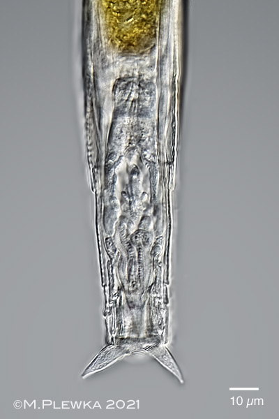 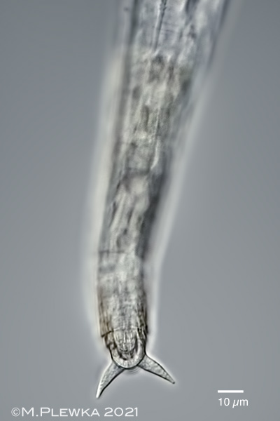 |
| 5. Rotaria tridens; foot; left: partially contracted specimen; focus plane on the footglands with partially granulated content. Right: expanded specimen; focus plane on the spurs with interspace (spur length (SL): 16µm |
| |
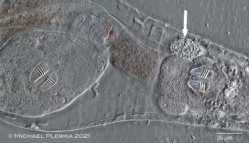 |
| 6. Rotaria tridens; specimen compressed by coverslide. The arrow points to one of the ?subcerebral? glands that is filled with fungal parasites. Like other Rotaria-species R. tridens is viviparous; on the left side of the image the trophi of the daughter can be seen, which have the same dental formula as the mother DF= 3/3) (2). Rami length (RaL): 24µm |
| |
 |
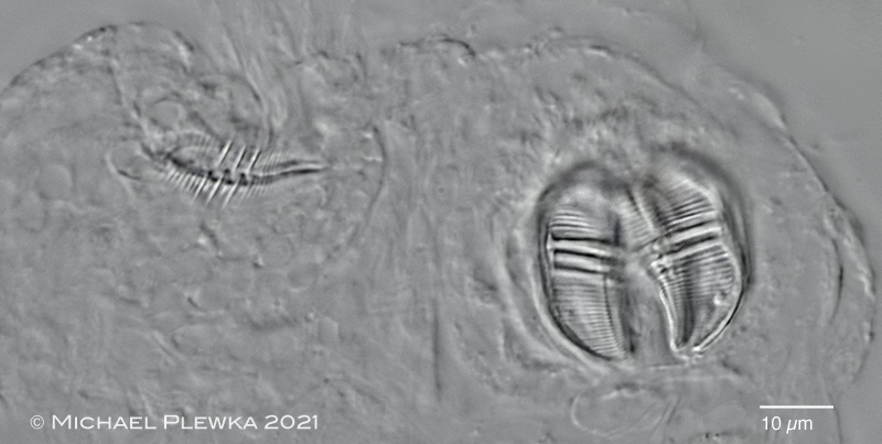
| 7. Rotaria tridens; in contrast to the above observation in several specimens it could be observed that dental formula of the trophi of the daughter (left) is different (DF: 3/2) from that of the mother (right, DF: 3/3). (2) |
| |
 |
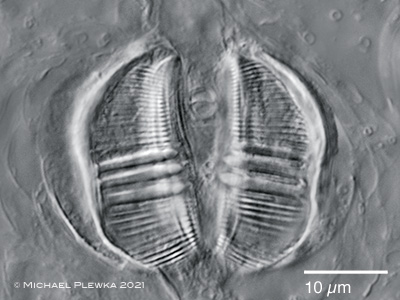
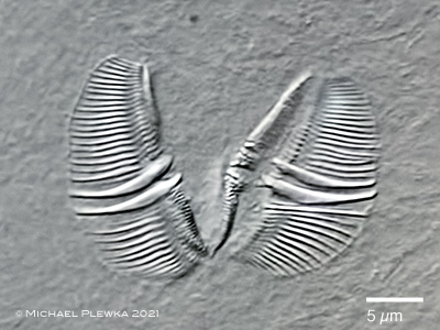
| 8. Rotaria tridens; trophi of another compressed specimen of the same morphotype. Trophi of daughter: left; trophi of mother: right. This image demonstrates that adult specimens of this species may have a dental formula of 3/2 (trophi on the right). The conclusion is that the DF: 3/2 of the trophi of the daughter in in the image (7) above (left) is the final stage of development. From the image here can also be concluded that the increase of the number of major teeth (mother DF: 3/2 >> daughter DF: 3/3) is possible. These observations give evidence that the the DF of bdelloid rotifers may vary, at least in this species. This has also the consequence that the identification of bdelloid species gets more complicated by the ambiguousness of the dental formula. (2) |
| |
 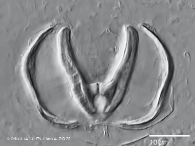 |
| 9. Rotaria tridens; trophi of adult specimen; Left: cephalic view; right: caudal view; ramus length (RaL): 23µm. |
| |
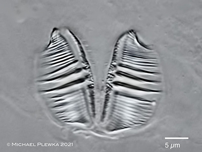  |
| 10. Rotaria tridens; 2 trophi of different embryos with different dental formulas of the same morphotype (number of nuclei of the vitellaria of the mother: 4). The manubria are only weakly developed at this developmental stage. (2) |
| |
| |
| Collection of all samples courtesy of Uli Drabiniok |
| |
|
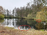
Location (1): Nature reserve NSG Heiliges Meer, NRW, Germany, "Fischteich" (location 17) (1); |
 |
| |
| Habitat(1): anoxic detritus, together with rhodobacteria, metopid ciliates and Mytilina trigona (1) |
| |
| Date (1): coll.: 13.11.2021; img.: 21.11.2021 (1); 24.11.20121 (2) |
| |
|
|
|
|
|
| |
|
|
|