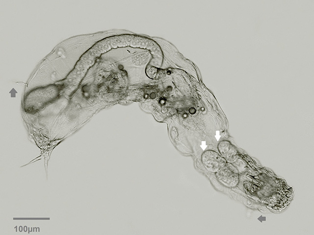
Tetrasiphon hydrocora, ventral view. Image taken without coverslip. Grey arrows: dorsal antenna, lateral antenna. White arrows: pair of gastric glands.
|
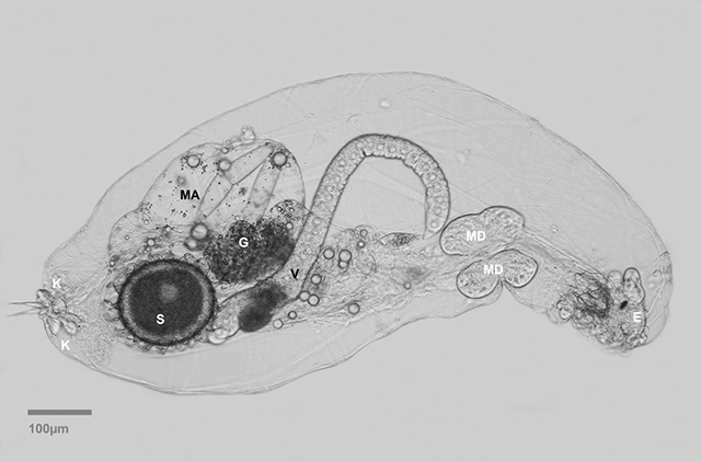 |
| Lateral view of a slightly compressed specimen. (E): eyespot, (MD): gastric glands, (V): vitellarium, (G): ring of glands between stomach and intestinum, (MA) intestinum, (S): resting egg, (K) adhesive glands. |
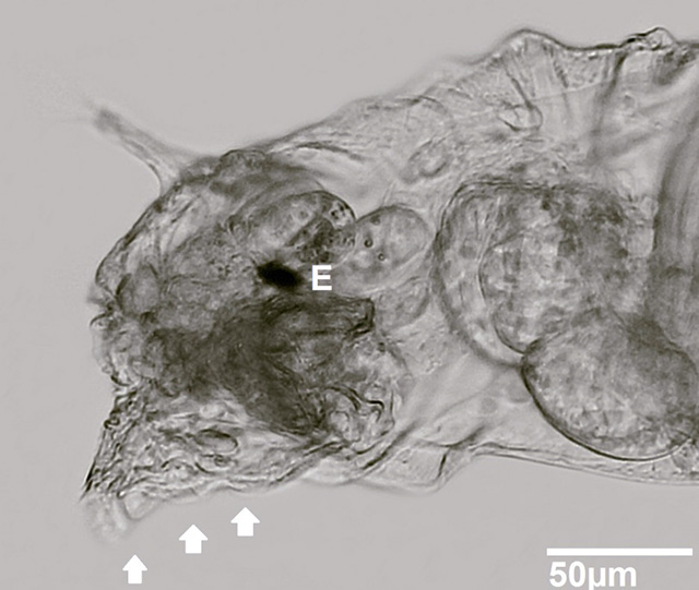 |
| Tetrasiphon hydrocora unfolding the corona (white arrows). (E): eyespot. |
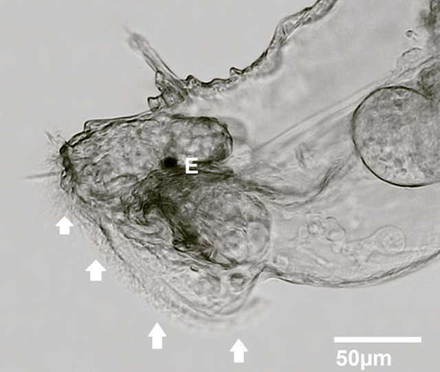 |
| Tetrasiphon hydrocora whirling. White arrows: corona. (E): eyespot. |
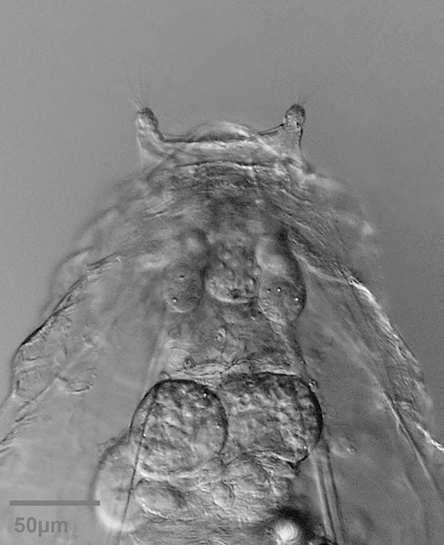 |
| Top view on the dorsal antennas. |
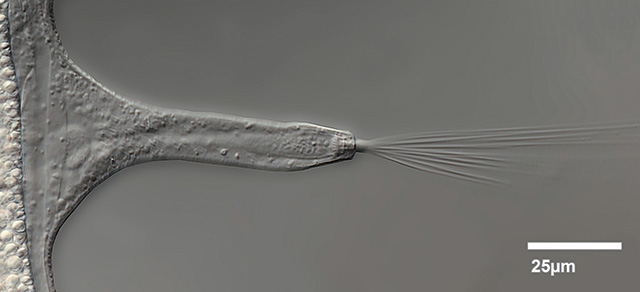 |
| Lateral antenna. |
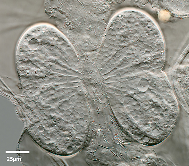 |
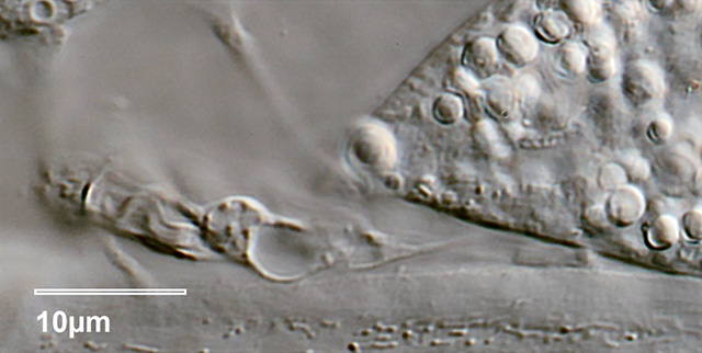
| Pair of reniform gastric glands. |
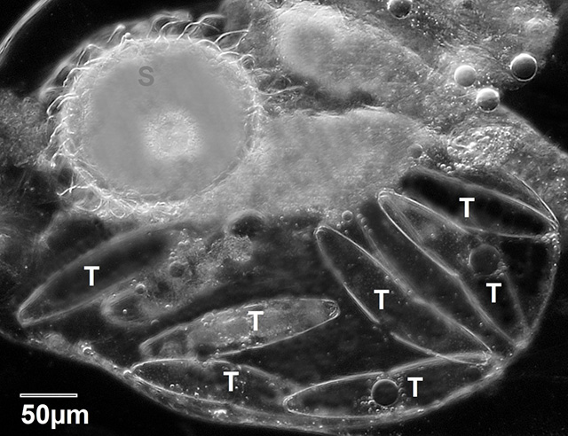 |
| Darkfield image of the intestinum. As described in literature, T. hydrocora is specialized in feeding desmids, in this case Tetmemorus granulatus (T). (S): resting egg. |
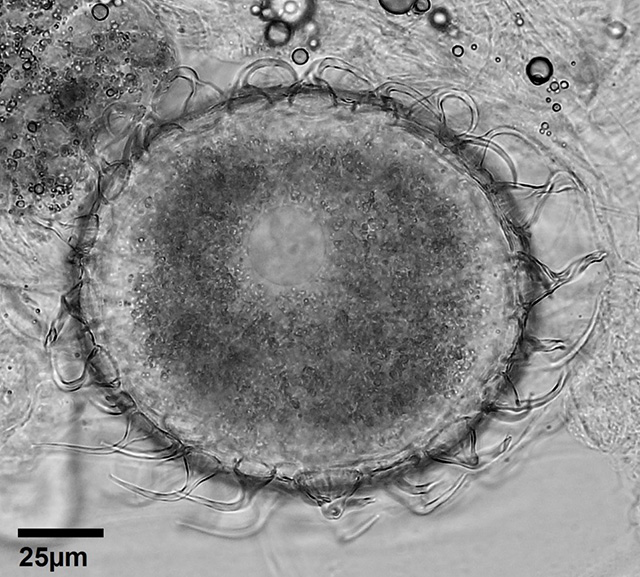 |
| Resting egg with helmet-shaped hairs. |
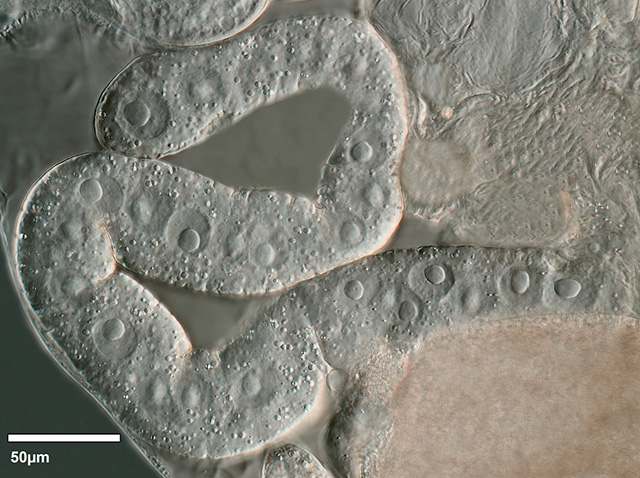 |
| Ribbon-like Vitellarium with approximately 30 nuclei. |
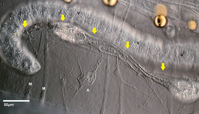 |
View on the nephridial system (yellow arrows). (T): terminal-cell.
(M): muscle fibres, (A): amoeboid tissue, (V): Vitellarium |
 |
Detail of the nephridial system: terminal-cell.
|
 |
| Detail of the nephridial system: cilia-cell. |
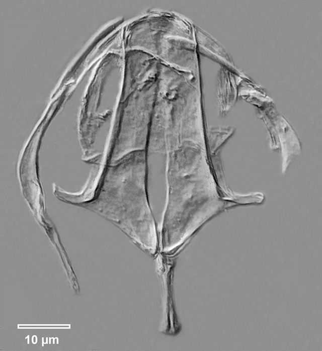 |
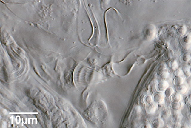
| Tetrasiphon hydrocora, trophi |
|
Location: Moor near lake Serrahn, Mecklenburg-Vorpommern/Germany
|
Habitat: Submersed moss , together with Beauchampiella eudactylota
About one specimen per 25 ml. Conductance-value: 20µS, pH: 5.3.
|
Date: collected 17.7.2015, images: 04.8.2015
|
All images courtesy of Richard Scholz.
|
|
|
|
|