| head of Philodinida |
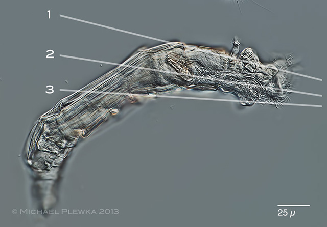
The head is the anterior part of a bdelloid rotifer |
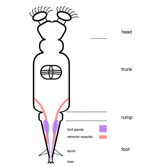 |
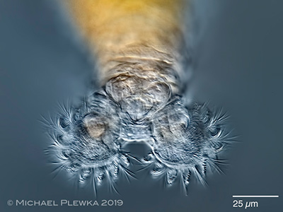
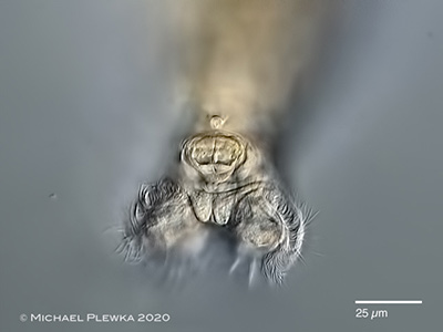
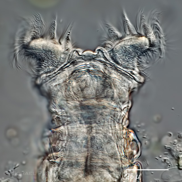
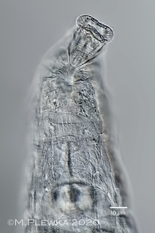
| Schematic diagram of the foot of a bdelloid rotifer |
| |
| |
 |
| |
 |
 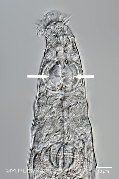 |
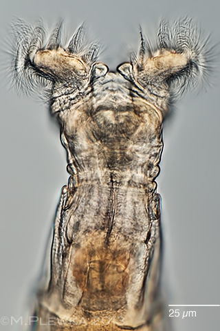
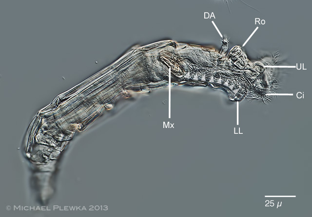
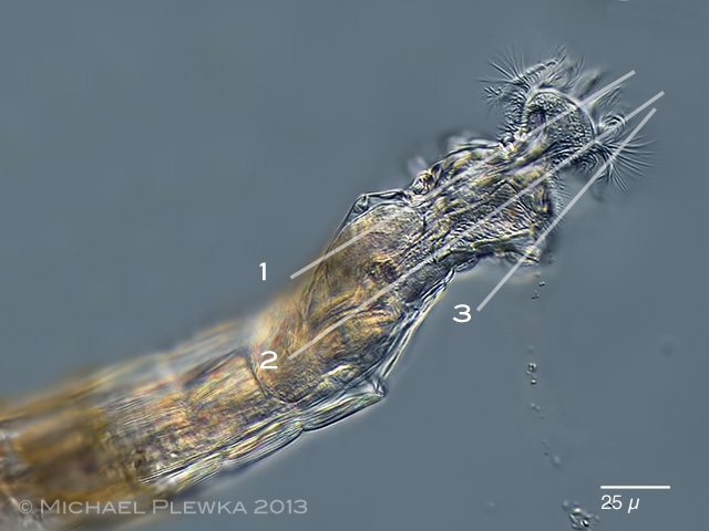
| Macrotrachela sp.5, the two images show the same specimen. Juxtaposition of specimen while whirling (left image) and while creeping (right). Left: head and neck; the dorsal antenna (DA) always inserts at the 1st neck pseudosegment (NS1). Ci: cingulum; DA: dorsal antenna; NS1-NS3: neck pseudosegments; Ro: rostrum (which is orientatd perpendicular to the longitudinal axis of the specimen); Tr: trochi; UL: upper lip. |
| The right image shows the anterior part of a creeping specimen. The rostrum is in the front of the head. Visible are the rostral cilia and the the invaginated trochal discs (arrows). |
 |
| Macrotrachela nana ligulata rotifer, whirling |
  |
| Philodina rotifer, whirling. |
  |
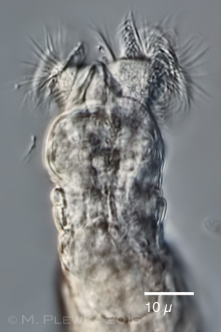
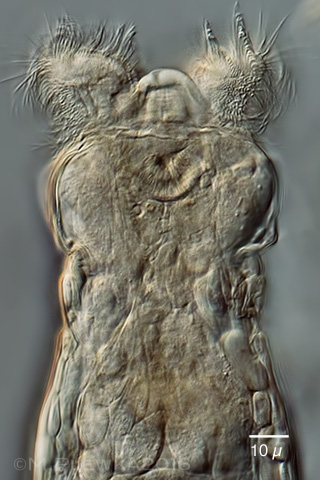
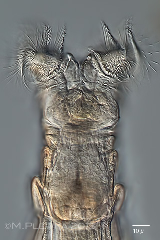
| Macrotrachela cf brachysoma rotifer, whirling. left/ right. Upper lip. |
 |
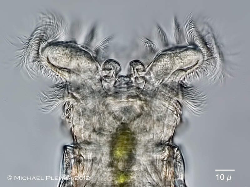
| Macrotrachela compacta rotifer, whirling. Upper lip. |
| |
 |
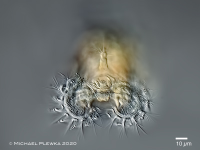
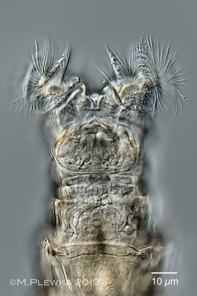
| Didymodactylos carnosus rotifer, whirling. Upper lip. |
| |
| |
| |
| |
 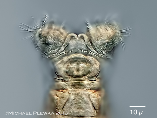  |
| Macrotrachela ehrenbergii rotifer, whirling. left/ right. Upper lip. |
 |
| Macrotrachela ehrenbergii rotifer, whirling. left/ right. Upper lip. |
| To get the best view on the toes the position of the rotifer should be "upside down", which happens very often when the rotifer is creeping. |
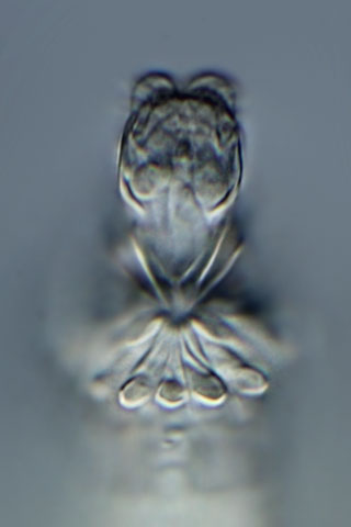  |
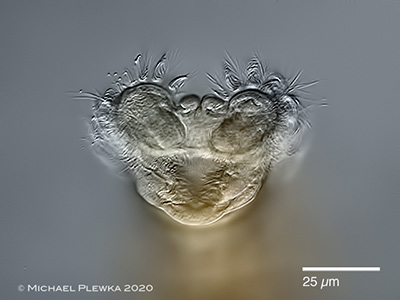
| left: Macrotrachela papillosa rotifer, creeping; mouth./ right: Macrotrachela brachysoma |
| bolsters |
  |
| |
| Macrotrachela magna left; Macrotrachela plicata right |
| |
 |
| |
 |
| |
| |
 |
| |
| |
| |
| |
| |
| |
| |
| |
| |
|
|
|
| |
| |