 |
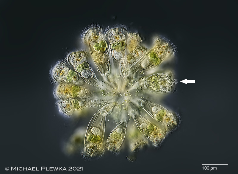
| (1) Conochilus hippocrepis, a pelagic rotifer. The arrow points to the two distinct lateral antennae, which separates this species from Conochilus unicornis. Colony from (3). |
| |
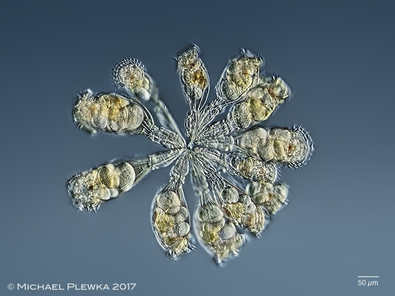 |
| (2) Conochilus hippocrepis, colony from (2). |
| |
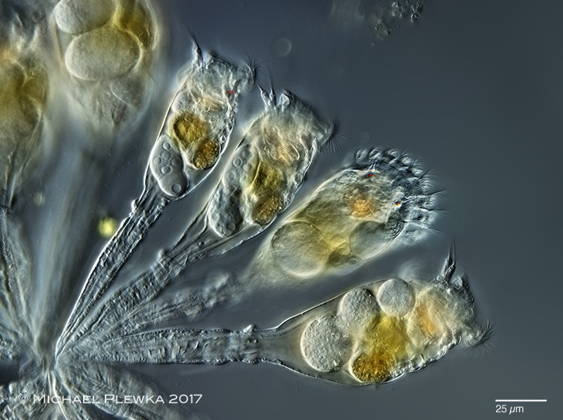 |
| (3) Conochilus hippocrepis, excerpt from colony from (2). |
| |
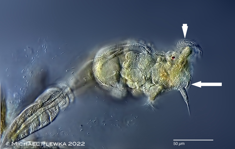 |
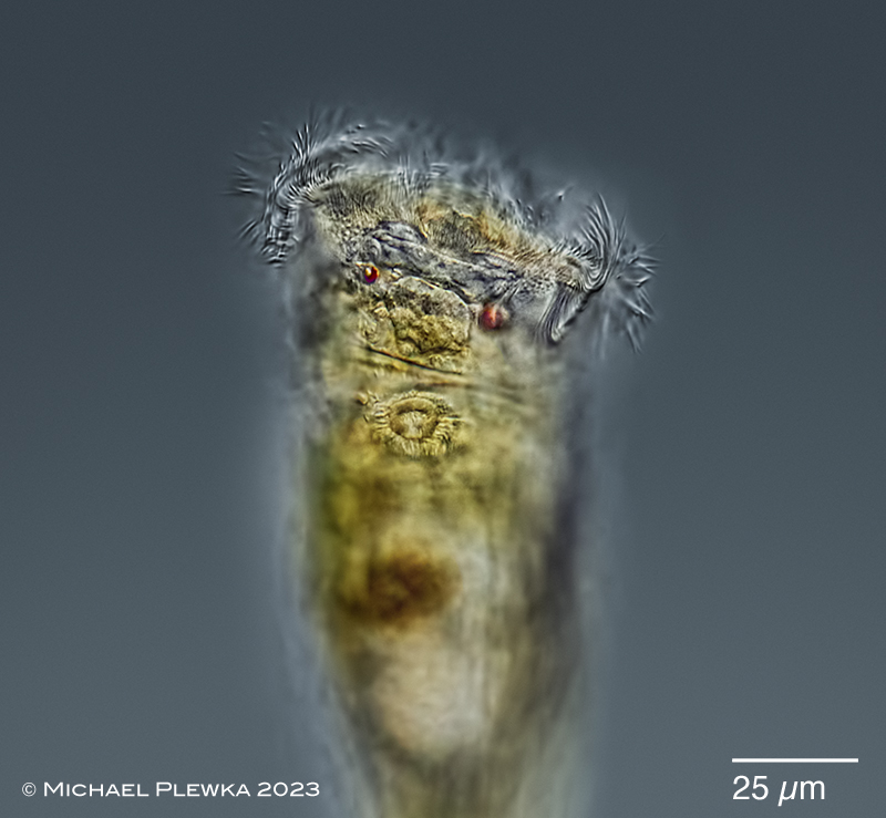
| (3b) Conochilus hippocrepis, specimen of colony from (5), lateral view. Optical longitudinal transect; the arrow points to the cone-shaped base of the lateral / ventral antennae; one of them is in focus in this image. This cone-shaped base is also the most ventral part of the cingulum, the cilia of which transport the diet to the mouth which located more dorsally. Also one of the red eyespots and the trochal cilia of the corona are visible. The shape of these cilia (arrowhead) suggests that the recovery stroke of the cilia is to the back, so the the effective stroke is forward, which causes the backward swimming of specimens that have (accidentely) detached from the colony. (5) |
| |
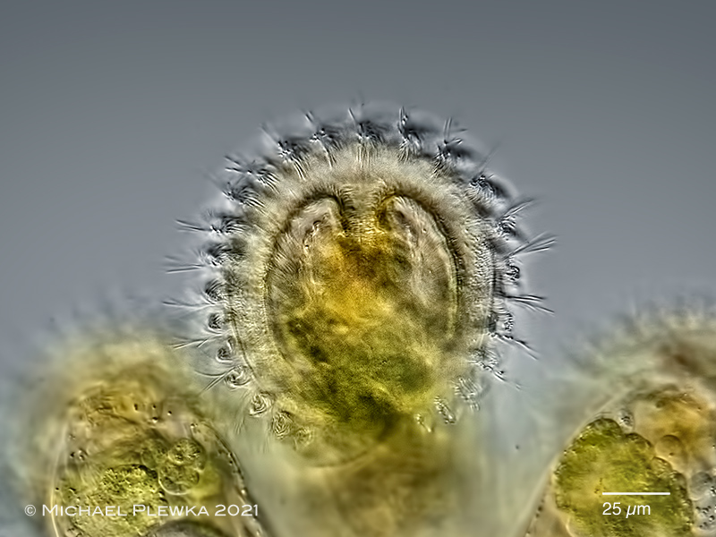 |
| (4) Conochilus hippocrepis, corona of specimen from (3). In contrast to the corona of the flosculariid rotifers Beauchampia, Floscularia, Limnias, Ptygura, Sinantherina, which have a closed circular corona, the corona of the Conochilidae is open, i.e.: horseshoe-like (see image below). The outer ciliated ring is the trochus; the inner ciliated ring widens in the middle to the buccal field of the oral cavity. |
| |
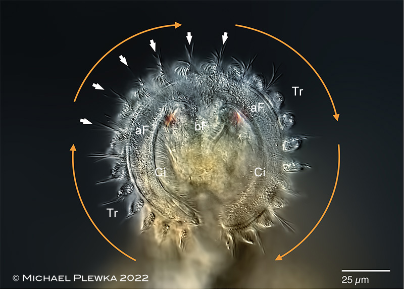 |
(5) Conochilus hippocrepis, corona of specimen from (4). In contrast to all other rotifers observed up to now (where the metachonous beating of the trochal cilia is counter-clockwise) the apparent "turning of the wheel" i.e. the metachronous movement of the trochal cilia of this species (genus??) is clockwise (orange arrows). What causes this different direction is not known. The whole "circle" (actually the ciliary wreath is interrupted at the ventral side) seems to be divided into 24 wave packets (some of which are marked by arrowheads).
Maybe this is also the reason for the "reverse" swimming of single specimens of Conochilus: foot is front; corona is back while swimming.
aF: apical field; bF: buccal field; Ci: cingulum; Tr: trochus. |
| |
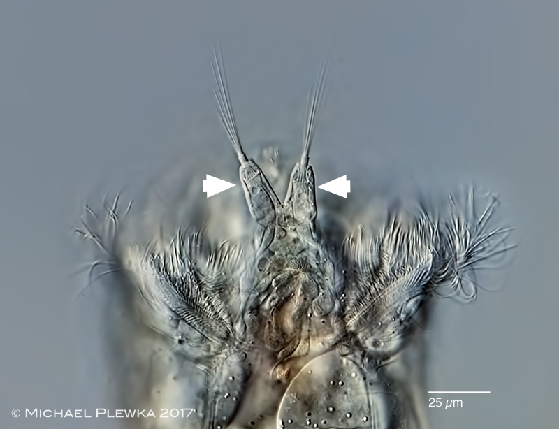 |
| (6) Conochilus hippocrepis, ventral view; in contrast to Conochilus unicornis this species has two more or less separated lateral antennas (2). |
| |
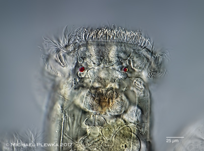 |
| (7) Conochilus hippocrepis, dorsal view; focus plane on the red eyespots.(2) |
|
| |
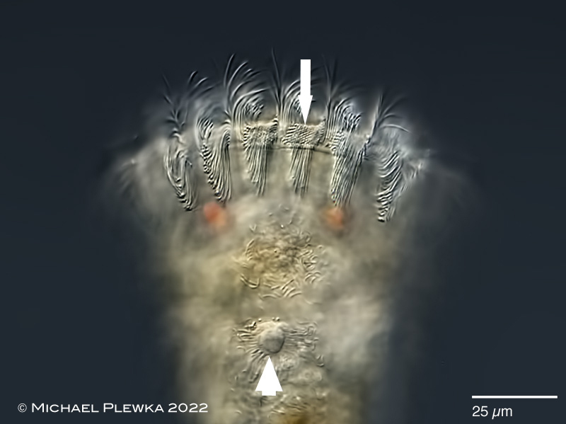 |
| (8) Conochilus hippocrepis, dorsal view; snapshot of the metachronal beating of the trochal cilia. 6 of the 24 wave packets are in focus here. The image also shows that the cilia are aranged in rows that are arranged at an angle of ≈ 30 degrees with reference to the trochus (arrow). Because the metachronal movement of the cilia is clockwise (when viewed anteriorly, see img. 5) and the effective stroke is in forward direction (see img. 3b) the metachronal coordination is dexioplectic. From observations with footage recorded at 180 fps it can be calculated that the speed of "rotation" is ≈ 40 rpm. (4) |
| |
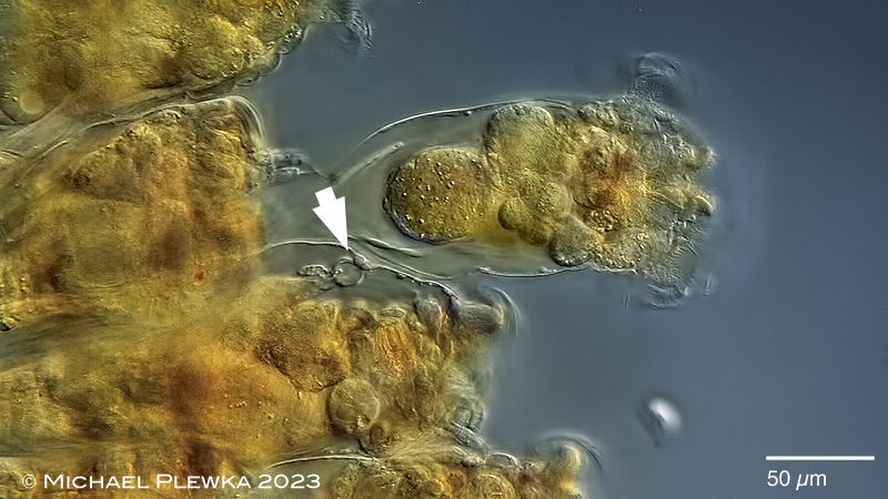 |
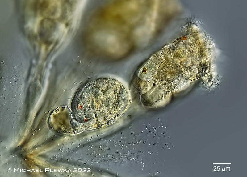
| (8b) Conochilus hippocrepis, part of a colony. The animal in the upper right part of the image is in dorsoventral view. The arrowhead points to the the end of the intestinum / bladder / anus of another animal in lateral view. This structure had been misidentified before as dorsal antenna in the image above (arrowhead). The same structure is also visible in the image below. (5) |
| |
 |
| (9) Conochilus hippocrepis, dorsal view of a specimen from; focal plane on the anus which is located near the head in this species; this had been misidentified as dorsal antenna before. (5) |
| |
 |
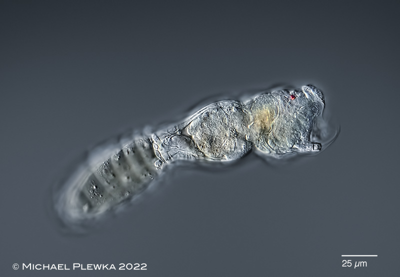
| (10) Conochilus hippocrepis, juvenile specimen, hatched 4 minutes before (4) |
| |
 |
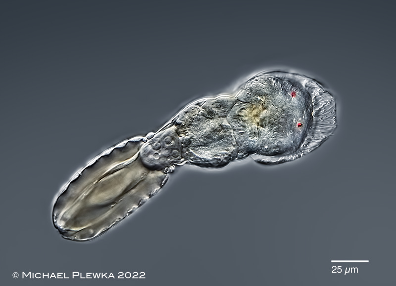
| (11) Conochilus hippocrepis, juvenile specimen (hatched 15 minutes before this image was photographed), lateral view, showing the position of the right lateral antenna in relation to the right eyespot. (4) |
| |
 |
| (12) Conochilus hippocrepis, same juvenile specimen, dorsoventral view. The cilia of the corona seem to be stil covered with a membrane from the egg shell. (4) |
| |
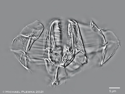 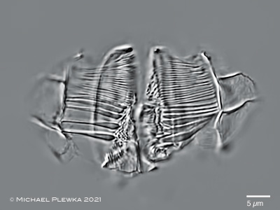 |
| (13) Conochilus hippocrepis, two aspects of the malleoramate trophi, different focal planes (3) |
| |
| |
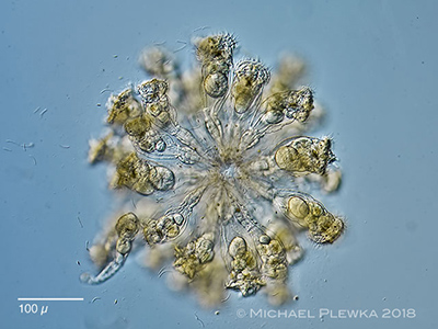 |
| (14) Conochilus hippocrepis, colony from (1) |
| |
| |
| |
|
|
| |
| |
| |
|
|
| |
| |
| |
| |
| |
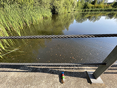
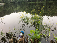
| Location: NSG Schwalm, Venekoten-See (1); NSG Heiliges Meer; Großes Heiliges Meer (2); Glör-Talsperre, NRW, Germany (3) |
| Habitat: Plankton (1;2); Plankton / between plants (3) |
| Date: 03.09.2011 (12); 21.04.2017 (2); 21.05.2021 (3) |
|
|
|