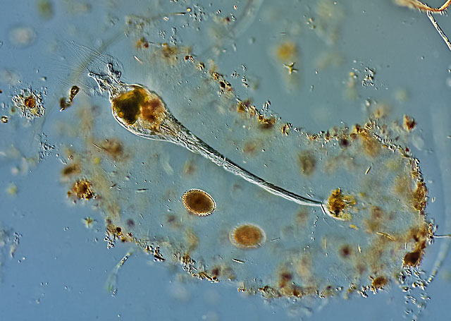
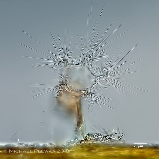
| Collotheca coronetta, total view; has a transparent mucilaginous sheath with enclosed detritus and bacteria. This tube is secreted by the integument, into which the animal can retract.Below the foot two resting eggs are recognizable. (1) |
| |
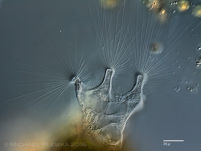 |
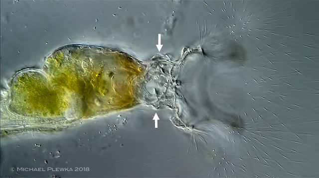
| Collotheca coronetta, corona. (1) |
| |
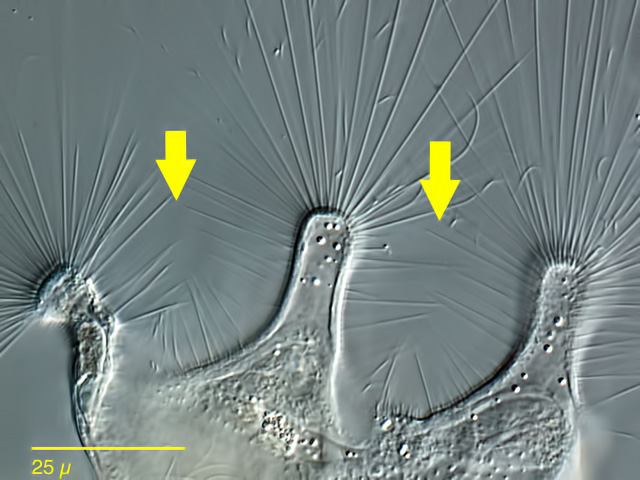 |
| Collotheca coronetta, a Collotheca-species with 5-lobed corona. The arrows point to the shorter cilia inserting between the lobes. This trait is in contrast to Collotheca ornata. (1) |
| |
| |
 |
| Collotheca coronetta coronetta, another specimen from (2) |
| |
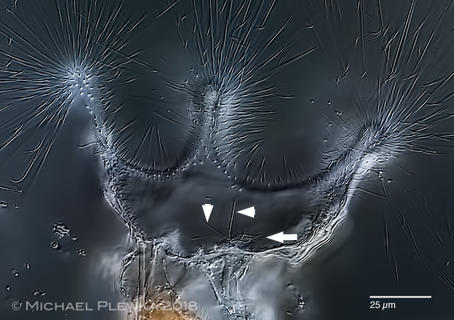 |
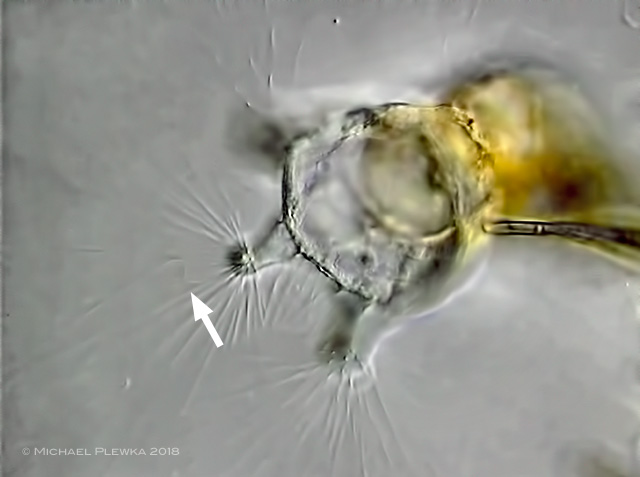
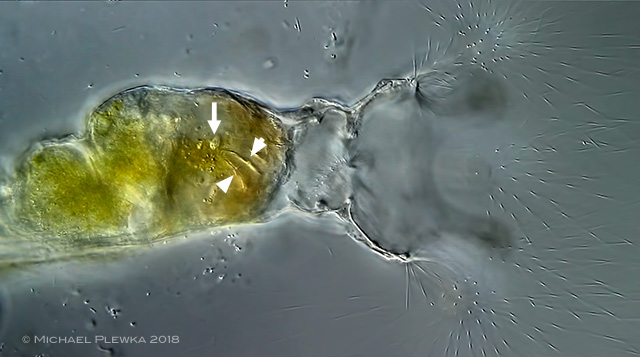
| Collotheca coronetta coronetta, crop of the above image. The arrowheads point to straight immobile cilia/ cirri, possibly with sensory function, while the arrow points to motile cilia which may transport the prey towards the vestibulum. |
| |
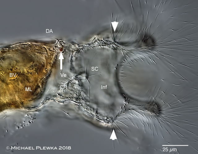 |
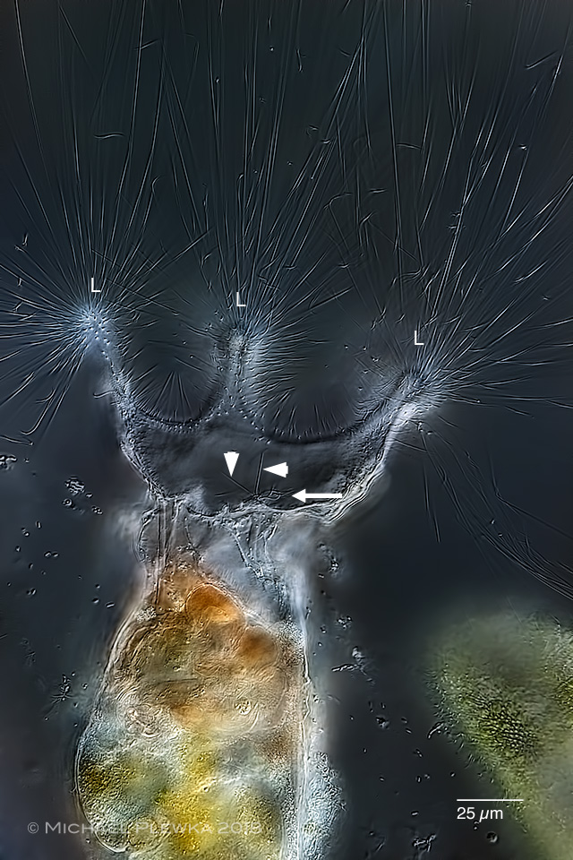
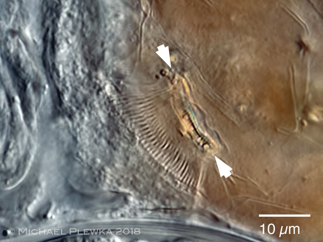
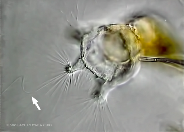
| Collotheca coronetta coronetta, another specimen from the same sample (2); optical longitudinal transect of the anterior part. The cilia marked by the arrowheads show that this specimen is C. coronetta coronetta. Visible is the dorsal antenna (DA); the arrow points to the eyespot. BV: pharynx/ buccal velum (part of) ;Inf: infundibulum; ML: mastax lumen/ proventriculus; SC: sensory cilia/ cirri; Ve: vestibulum. |
| |
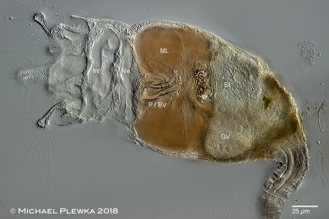 |
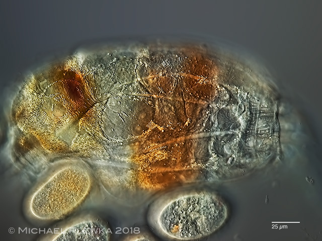
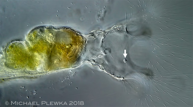
| Collotheca coronetta coronetta, another specimen from the same sample (2); the course of prey goes from left to right. Here, the pharynx or the undulating membrane of the pharynx (P / BV) can be seen more clearly. The reddish-brown coloration of the liquid content of the mastax lumen suggests that there is already a pre-digestion or (at least microscopically visible) immobilization of moving prey organisms. At the end of the proventriculus/mastax lumen (i.e., right) are the hard parts (trophi) with the muscles. There, the food is either minced, which works for example on prey organisms with "soft" cell wall (e.g. euglenoid flagellates), or only that part of the prey organisms is carried further to the right in the actual stomach (St), which really fits through, e.g. in prey organisms with a silicious cell wall (diatoms) only the lipid droplets are passed through and the cell walls are deposited in the mastax lumen. See Collotheca edenta as example. GV: germovetellarium; Inf: infundibulum; P/BV: pharynx with buccal velum; St: stomach; Tr: trophi; Ve: vestibulum. |
| |
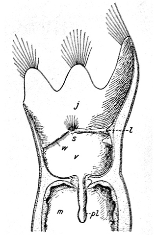 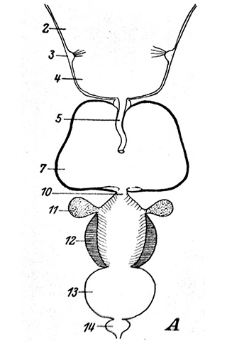 |
Head and digestive tract of collothecid rotifers; left image: Collotheca sp. ; right image: Stephanoceros fimbriatus; both images from Remane (1929). In principle, the morphology and function of the organs of the head region and the digestive system should be identical and thus homologous in the genera Collotheca and Stephanoceros. Now the caption to these pictures, however, is interesting:
left picture (excerpt) : "...j Infundibulum; l: Diaphragma; ...s: Sinnesorgan des Schlundrohrs, (sog. Pharyngealsinnesorgan)..."
right image (excerpt): "....2 Vestibulum; 3 Pharyngealsinnerorgan; 4 Infundibulum..."
So in the left picture, the infundibulum is the 1st section of the digestive tract, followed by the vestibulum, which opens into the mastax lumen via the pharyngeal tube. In the right picture it is the other way round: the vestibulum is the 1st section, the infundibulum is the 2nd section.
Thus there is a contradiction in the designation of the organs.
Both illustrations served as models for later authors; for example, in the book of Koste (1978) there is an illustration of Collotheca trilobata which takes the designation from the left picture; in a rotifer chapter of of DeSmet / Fontaneto (2015) the designation of the right picture is taken over.
This is all the more regrettable as in other works by Hochberg et al. (2018) l the entire "head" area is referred to as the infundibulum, for whatever reason.
|
| |
 |
| Collotheca coronetta coronetta, same specimen as above, detail of the pharynx. In this image only a part of the pharynx is recognizable. One recognizes parallel running cilia, which together form a movable plane. The arrowheads mark an area where the membrane kinks and therefore becomes visible in the optical cross section. This undulating membrane appears to be actively involved in the transport of prey. (2) |
| |
 |
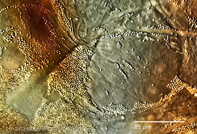
| Collotheca coronetta coronetta, top view of the body or the integument. You can see some bright longitudinal muscles. In the body fluid of Collotheca different amounts of granules can be found, depending on age. These are constantly being washed around in the pseudocoel by altering the volume ratios whenever the animal moves. What is the function of the granules, is probably not known. In some rotifers, e.g. in Asplanchna, freely moving cells (amoebocytes) have been observed very early in the pseudocoel.
These amebocytes also contain vacuoles that may contain bacteria or other bodies. The vacuoles have been assigned the function of serving - similar to amoeboid mobile cells in mammals (macrophages) - the immune system. Collotheca suggests that homologous but immovable cells take up these granules. These cells are called excretophors. (2) |
| |
 |
Collotheca coronetta coronetta, crop of the above image. Excretophores with granules. (2) |
| |
 |
| Collotheca coronetta coronetta, top view of the corona of C. coronetta. In one sample, I was lucky that a critter was almost perpendicular to the plane of the microslide. The photo shows the spatial orientation of the cilia, which corresponds to some of the drawings, as they can be found in KOSTE (1978). While in the typical side view (as shown in the first picture of this page) one can get the impression that the cilia are a more or less impenetrable thicket for prey organisms, this figure makes it clear that this assumption is false. Although the depth of field is low in this recording, it also shows while focusing through the specimen that the long cilia are mainly directed to the outside. A certain proportion of short cilia is aligned towards the center, so that in the center of the corona is a virtually unhindered entry and exit for prey organisms. At least that's the case with C. coronetta. In this respect, it is imperative to examine the morphology of the respective species in order to be able to make statements about the prey. (2) |
| |
 |
 |
| Collotheca coronetta coronetta, two stills from a video from the same specimen as above. The arrows mark a cilia over which a bend similar to a wave propagates from the base, i.e. from the lobe to the top. Such waves can be observed very frequently in the corona, without any cause (i.e., a prey organism) being recognizable. A similar wave also occurs on a rope when it is mechanically excited at one end. The conclusion is that this wave movement of the cilia is caused by contractile elements in the lobe. Such contractile elements at the base of cilia are well established for other rotifer species (e.g., Trichocerca). (2) |
| |
 |
Collotheca coronetta coronetta, 3 still images from a video of C. coronetta showing the uptake of a heterotrophic flagellate. Image 1:
A heterotrophic flagellate is actively moving its scourge into and around Collotheca's mouth / drogue. Sporadically, movements of the short cilia between the praise can be observed for fractions of a second. The moving cilia on the papillae at the base of the vestibulum are in constant motion. The flagellate moves back and forth in the vestibulum, briefly out again. After a few seconds, it moves into the infundibulum, while the cilia row (CR)are constantly moving (so it can be assumed that these cilia at least support the transport of the prey.. |
| |
 |
Collotheca coronetta coronetta, Img 2:
Collotheca's body then contracts in the area of ??the vestibule, so that the diameter of the animal at this point is smaller (no shortening in the longitudinal axis!), The pharyngeal area is quickly pushed forward and the flagellate is constantly moving the pharynx / undulating membrane in the Mastax lumen / Proventriculus transports. The long cilia on the lobes do not change. |
| |
 |
Collotheca coronetta coronetta, Img 3:
Visible is the undulating membrane of the pharynx (arrowsheads) as well as the uptaken flagellate (arrow). The prey in the mastax lumen / proventriculus is constantly moved by the movement of the pharynx back and forth. (Not seen in this video, but I've often seen elsewhere - flagellates in particular - the beating of the flagella for a few minutes in the mastax lumen / proventriculus or the metabol movement before the movement ceases). Conclusion: in this species Collotheca coronetta, the long cilia have contributed to this entire "prey catching" in no apparent way; the function as trap is not detectable. As already indicated: also also in the case of C. dentata the prey actively moves into the vestibulum. |
| |
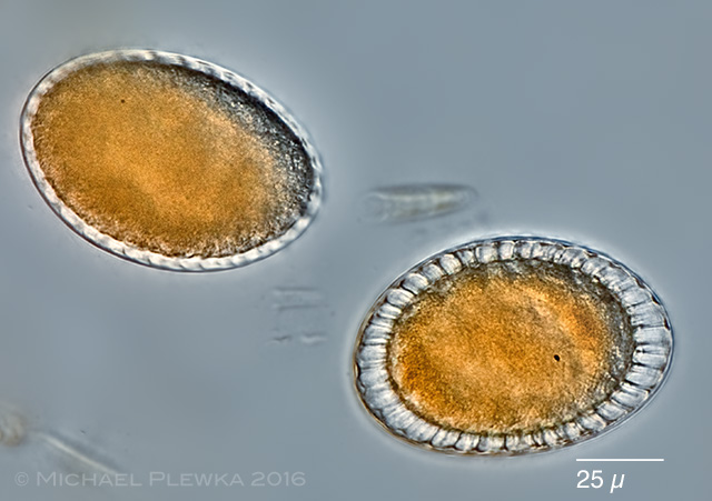 |
| Collotheca coronetta, two resting eggs. (1) |
| |
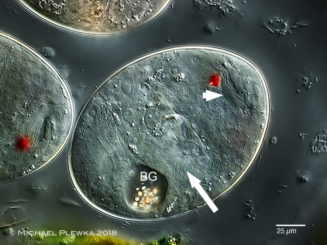 |
| Collotheca coronetta, amictig eggs. In the egg in the center odf the image the red eyespot and the birefringent granules (BG) are visible. The arrow and arrowhead point to two ciliated areas, The one marked by arrowhead near the eyespot may show the cilia of the future larval corona, whereas the cilia marked by the arrow may develop into the future infundibulum (see image below). (2) |
| |
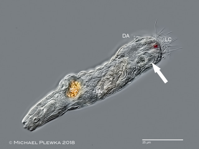 |
| Collotheca coronetta, juvenile specimen, swimming, lateral view. From the juvenile to the adult stage the specimen undergoes a metamorphosis. The larval corona (LC) does not develop into the "corona" of the adult stage, but will reduce in size. In this larval stage the cilia of the infundibulum (If) are already visible. This infundibulum is the "corona" of the adult stage. BG: birefringent granules; DA: dorsal antenna; F: foot; Mx mastax. |
| |
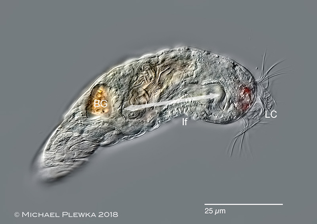 |
| Collotheca coronetta, same specimen. The double arrow marks the area of the infundibulum. |
| |
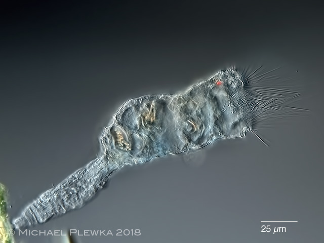 |
| Collotheca coronetta, juvenile specimen, 12 hours later, already attached to the substrate (alga). The birefringent granules in the intestinum are still visible. |
| |
| |
| Location: NSG Heiliges Meer, near Erdfallsee (1); Mariaveen near Griendsveen/ NL, pond with Utricularia sp. (2). |
| Habitat: periphyton: on the branches of Utricularia sp. (1); (2); sympatric with Collotheca heptabrachiata (1) |
| Datum: 15.05.2011 (1); 20.11.2018 (2) |
|
|