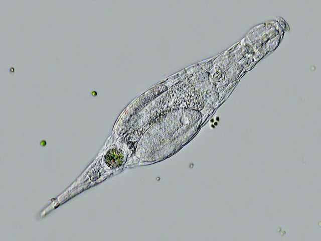
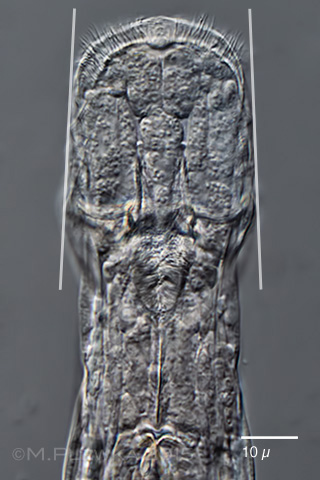
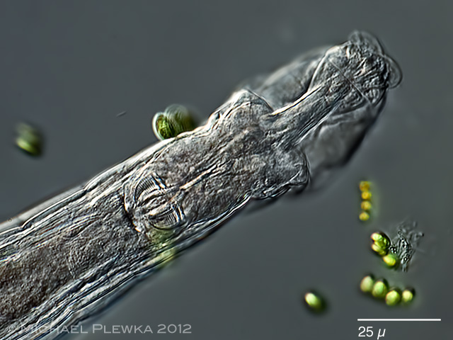
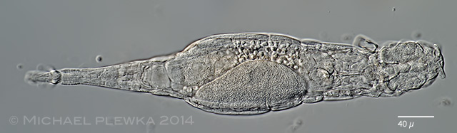
| Adineta vaga minor is a very common bdelloid found in many samples from soil and mosses in our region. It often occurs together with other Adineta species like A. vaga major or A. steineri. (1) In contrast to A. vaga major the transition between the trunk and first rump segment is more or less continuous and the first rump segment is not swollen. |
|
|
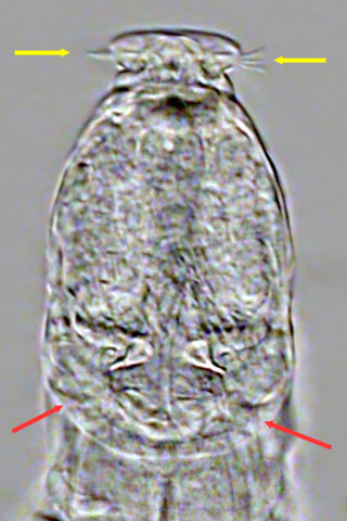
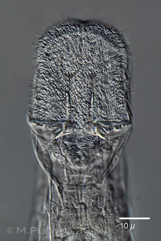
| Adineta vaga minor: head in two different focal planes, ventral view. The ventral side of the head is covered with cilia (ciliary field)which accomplish the moving of the rotifer. left image: bristles at the lamella of the rostrum (yellow arrows). The red arrows point to the cingulum. Right image: the arrows point to the 4 or 5 "U-hooks" on each side that act like rakes by scraping over the surface and gathering bacteria. (1) |
 |
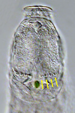
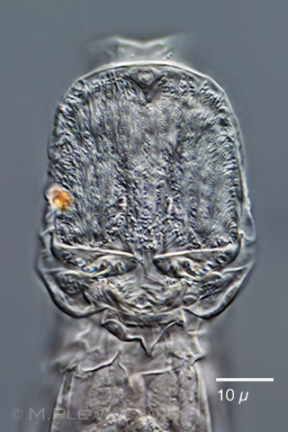
| Adineta vaga minor, this DIC-image of a specimen vital-stained with Neutral Red shows the ventral view of the cuticularized prehensile apparatus (rake apparatus). The prehensile apparatus is used to scrape off bacteria from the substrate. It comprises two rakes located at the posterior part of the ciliary field. Each rake bears a certain number (which seems to be species-specific) of denticles (labelled with arrows in the right rake) which are reinforced at their base with a material that is resistant against SodiumHypochlorite (NaOCl). When dissolving the specimen to view the trophi these reinforcing structures will remain visible as so-called U-hooks (arrowheads). So in Adineta minor there are 5 U-Hooks and 6 denticles in each rake. |
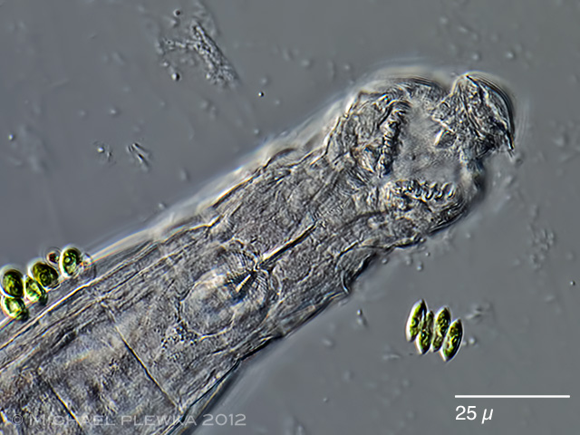 |
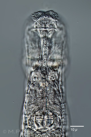
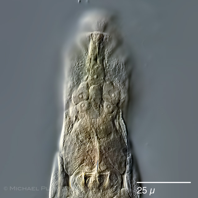
| Adineta vaga: another specimen, focus plane on the ciliary field and the rakes.(2) |
 |
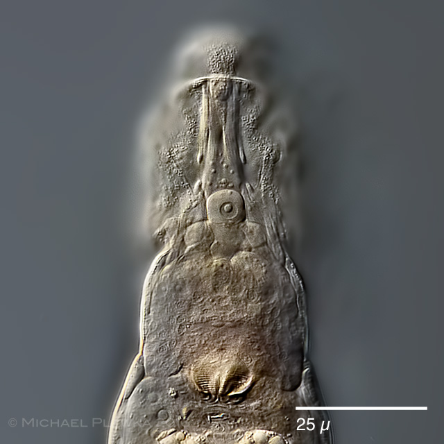
| Adineta vaga: same specimen, focus plane on the dorsal side of the head.(2) |
|
|
|
| Adineta vaga minor?, series of 4 optical sections through the head from dorsal to ventral side. Upper left: focus plane on the rostrum and nerve/ muscle cells. Upper right: focus plane on the hypodermic cells. Lower left: focus plane on the ciliary field. Lower right: focus plane on the rake apparatus. (4) |
 |
 |
 |
| Adineta vaga minor, series of 3 optical sections through the head from dorsal to ventral side; the images show the hypodermic cells with their nuclei. |
 |
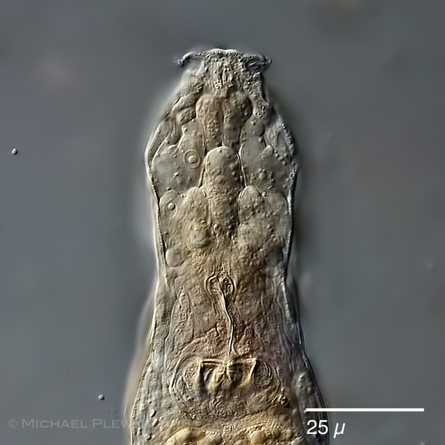
| Adineta vaga minor, specimen from (3). |
 |
| Adineta vaga minor, same specimen from (3) as above, focus plane on the ciliary field and rake apparatus. |
 |
| Adineta vaga minor, crop of the above image showing the 5 U-hooks. |
| Location: Gevelsberg, Stefansbachtal Schulzentrum, Gruenes Klassenzimmer (1) Bonampak, Mexico (3); Hattingen Oberstueter (4),(5) |
| Habitat: Moss on tree (Salix) (1,3); moss on roof (4); moss on sandstone (5) |
| Date: 17.8. 2008 (1) 02.11.2014 (4); 6.02.2016 (5) |