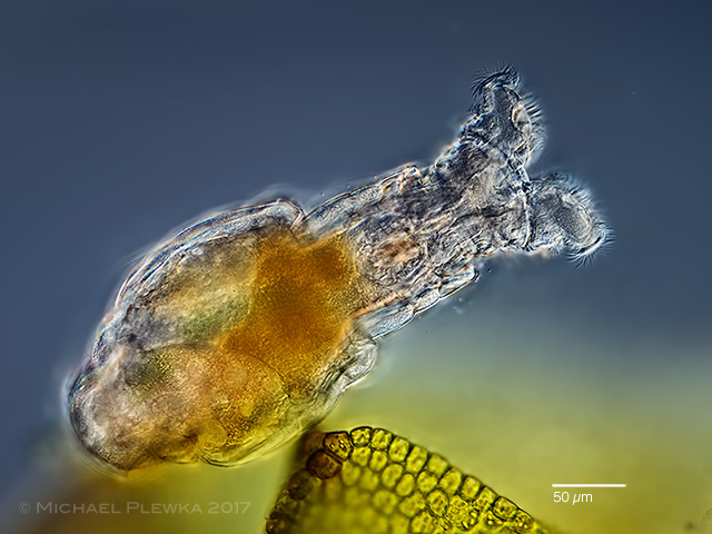
| Mniobia russeola; specimen living on Frullania dilatata. In contrast to Mniobia symbiotica or Habrotrocha annulata, which both live inside the lobules of Frullania the specimens from this sample were found between the leafs of the liverwort.(3) |
| |
 |
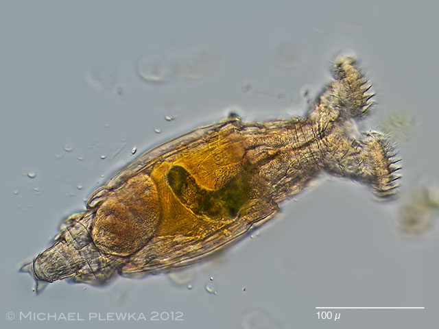
| Mniobia russeola, whirling, dorsoventral view; specimen from (1). A part of the images shown here on this page have been mismatched before as Mniobia magna, but the proportions of the head ( the head of this species is more conical than that of M. magna and the sulcus (= interspace between the trochal discs) is very wide) speak for M. russeola. |
| |
 |
| Mniobia russeola, another specimen from (2). Wide corona, narrow head. Focus plane on the dark green stomach lumen. |
| |
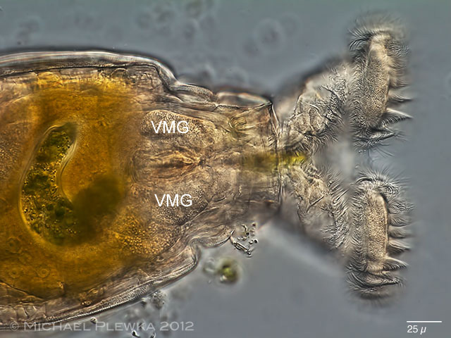 |
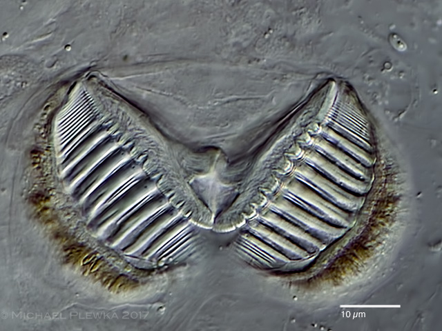
| Mniobia russeola, focus plane on the ventral side of the trochal discs. VMG: ventral mastacitory glands. (2) |
| |
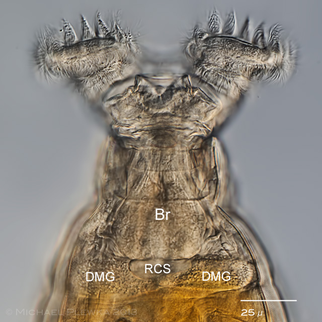 |
| Mniobia russeola, head region of the same specimen from (2). Focus plane on the dorsal side showing the pear-shaped brain (Br; some of the nuclei of the nerve cells are visible); DMG: dorsal mastacitory glands; RCS: retrocerebral sac. |
| |
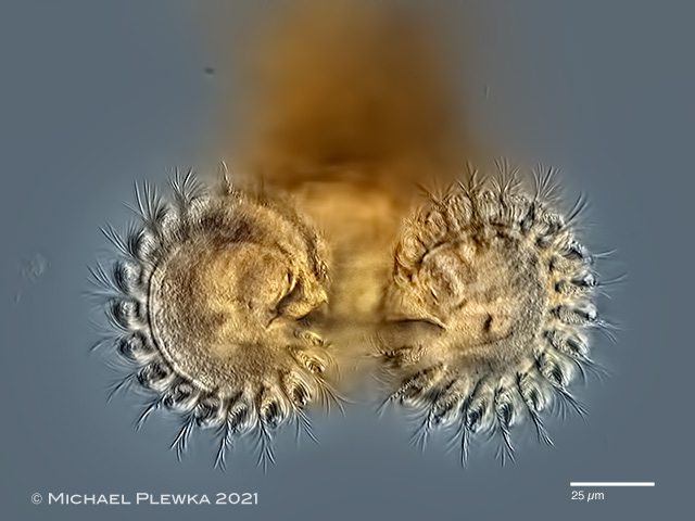 |
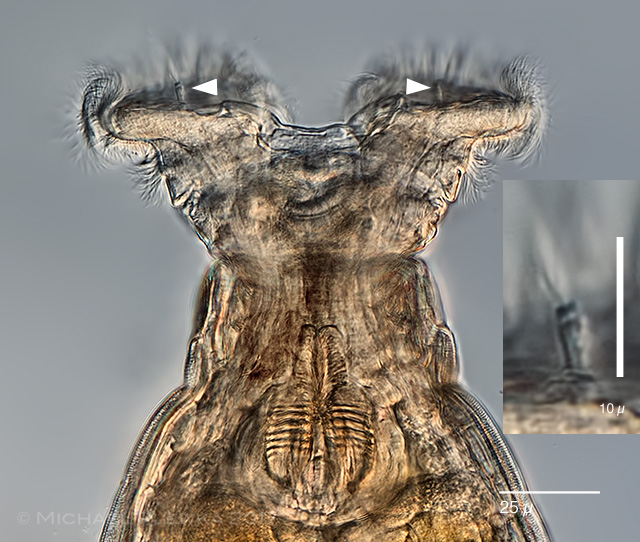
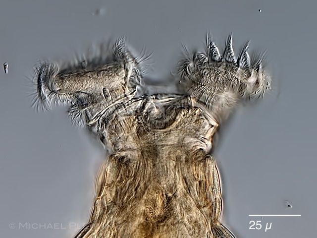
| Mniobia russeola, corona of whirling specmen, frontal view. (4) |
| |
 |
| Mniobia russeola, from the bent form of the body one can imagine the leech-like movement of the bdelloid rotifers. (15.11.2012) |
| |
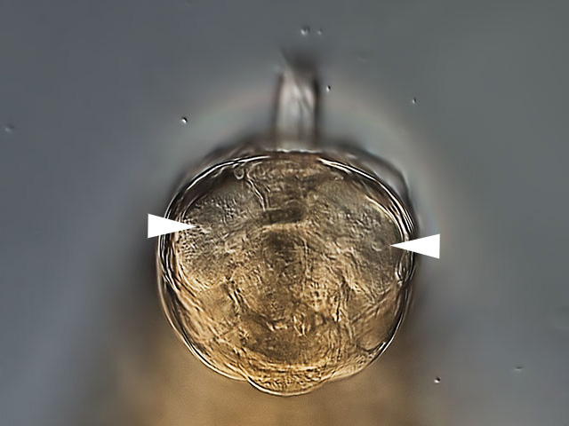 |
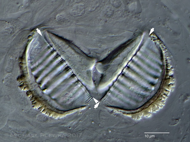
| Mniobia russeola, crop of the above image. The papillae on the trochal discs are visble(arrowheads). They bear sensory cilia (see image below). |
| |
 |
| Mniobia russeola; the arrowheads point to the sensory organs on the trochal discs. The insert (crop of the main image) demonstrates that the sensory organ is comprised of a papilla with several cilia. Also visible in the main image are the muscles prior to the mastax. (1) |
| |
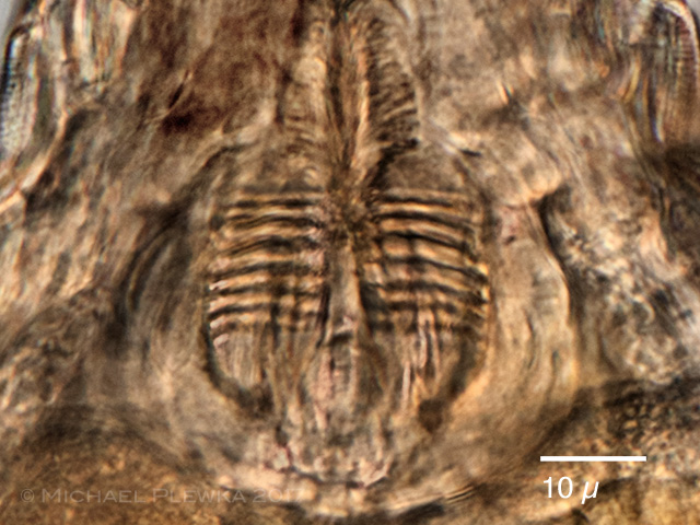 |
| Mniobia russeola; crop of the above image, mastax and trophi. (1) |
| |
 |
| Mniobia russeola; same specimen, focus plane on the upper lip. (1) |
| |
|
|
|
|
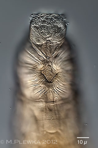
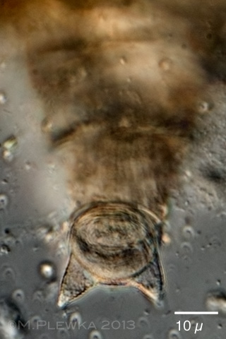
| Mniobia russeola; left image: anterior part of creeping specimen, focus plane on the rostrum lamella (2). Right image: foot with spurs and adhesive plate (1) |
| |
 |
 |
| Mniobia russeola; two images of the ramate trophi; dental formula: 8+1/9; rami length (RaL): 32µm. Upper image: cephalic view; lower image: caudal view. Are the lines (each is marked by two arrowheads) the reason why De Koning stated that the teeth cover only one half of the unci plates? (3) |
| |
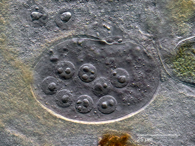 |
| Mniobia russeola; germovitellarium with nuclei. (3) |
| |
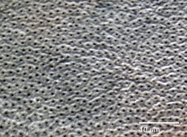 |
| Mniobia russeola; integument with pores (oil immersion; Zeiss Planapo 100x 1.4). In contrast to Mniobia scarlatina the integument has no protuberances, but tiny elevations which seem to form rows which is in contrast to the morphotype of Mniobia magna. |
| |
| Location: Eifel, Germany (1) NSG Heiliges Meer, Germany (2); Obfelden, Switzerland, forest (450moh) (3); Südtirol, Italy; Schrüttensee, |
| Habitat: beech litter (Fagus)(1); pond with Sphagnum (2); Abies alba, bark , on Frullania dilatata (3); moss (3) |
| Date: 07.10.2013 (1); 15.11.2012 (2); coll.: 12.12.2017, img.: 20-22.12.2017 (3); coll.: 08.2020; img: 05.01.2021 (4). |
| Collection of sample No. (3) by courtesy of A. Bueschlen, swissbryophytes |
| Collection of sample No. (4) by courtesy of Pit Staedler, Gevelsberg. |
| |
|
|