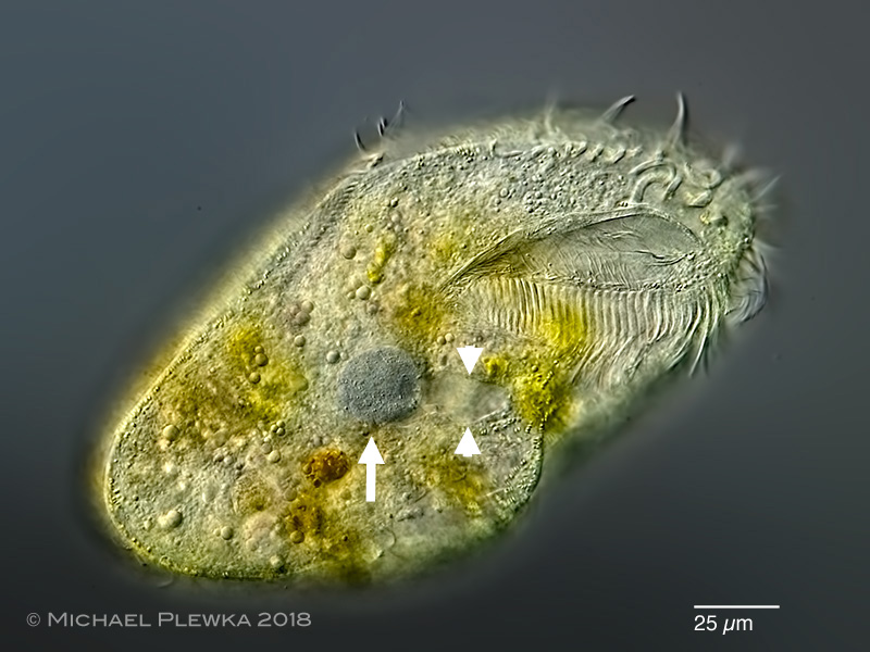
| Podophya urostylae; the arrow points to this ectoparasitic suctorian, which has infested the hypotrich ciliate Urostyla grandis (which is the swimming ciliate in this image). P. urostylae may be misinterpreted as the macronucleus of the host. The arrowheads point to the invagination / canal which connects the parasite to the environment. |
| |
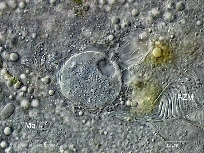 |
| Podophya urostylae; some of the tentacles protruding into the invagination (In) are visible in this image. (AZM: adororal zone of membranelles of the host; Ma: macronucleus of the host). |
| |
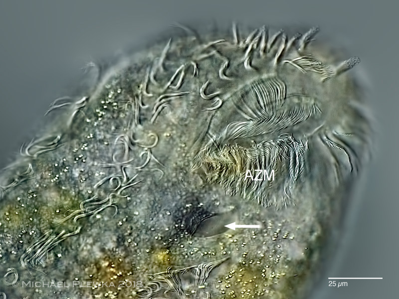 |
| Urostyla grandis; the arrow points to the opening of the invagination in the cortex, which goes further into the center of the cell, where P. uroystyla is located. The ciliated swarmer of P. urostylae adhereres to the cortex and causes the invagination. It then looses the cilia, penetrates into the host and starts to suck the host with the tentacles. The invagination keeps open as long as the parasite is inside. According to the literature up to 50 parasites have been observed in a single host! |
| |
| Identification courtesy of Dr. Martin Kreutz, Konstanz |
| |
| Location: Mariaveen near Griendtsveen/ NL, pond |
| Habitat: ectoparasitic on / in Urostyla grandis (which orrured in the detritus) |
| Date: 18.11.2018 |
| |
|
| |
| |
|