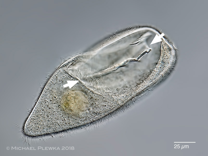
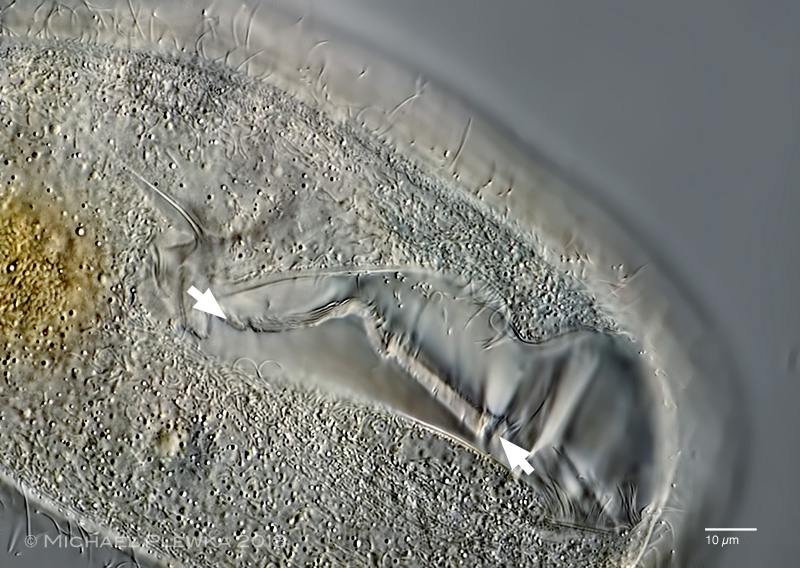
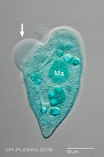
Turaniella vitrea; focus plane on the conspicuous oral apparatus with (one of?) the adoral mebranelles (arrows). (2)
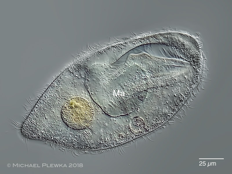
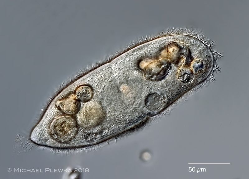
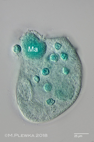
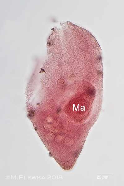
Turaniella vitrea; focus plane on the macronucleus (Ma). (2)
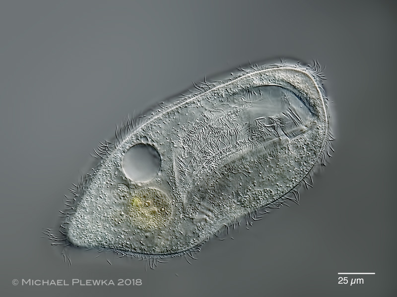
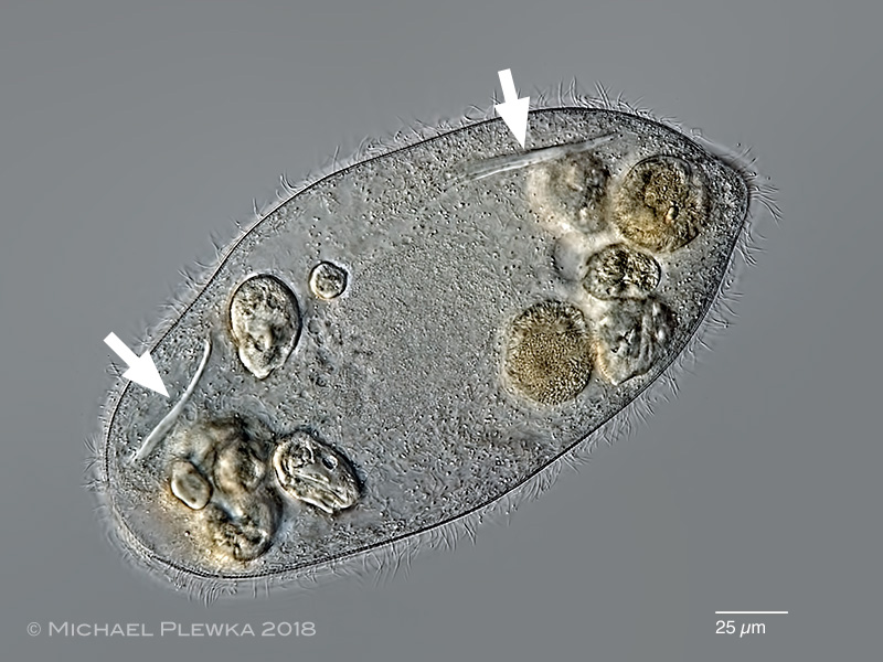
Turaniella vitrea; focus plane on the contractile vacuole and the cilia of the ?undulating membrane?
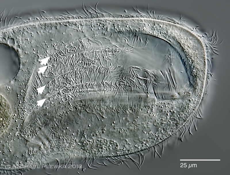
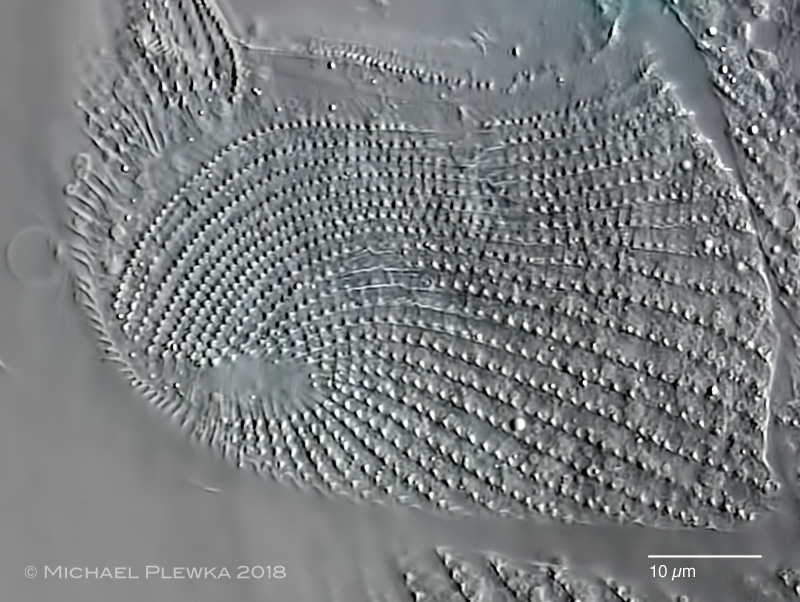
Turaniella vitrea; crop of the above image. Oral apparatus; different focus plane. At least four rows of cilia are recognizable in the oral apparatus.(3)
Turaniella vitrea; the arrows mark (one of?) the adoral membranelles of the oral apparatus. (3)
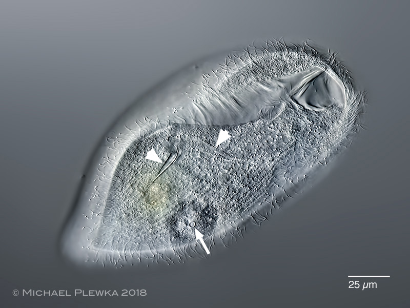
Turaniella vitrea; focus plane on the cortex. The arrowheads point to two unknown structures. The arrow points to the excretion pore of the contractile vacuole.
Turaniella vitrea; dividing specimen, slightly compressed by the coverslide. During division the undulating membranes and the opening of the oral apparatus are not visible, but the membranelle which is already replicated. (The posterior end is in the right upper corner in this image) (1)
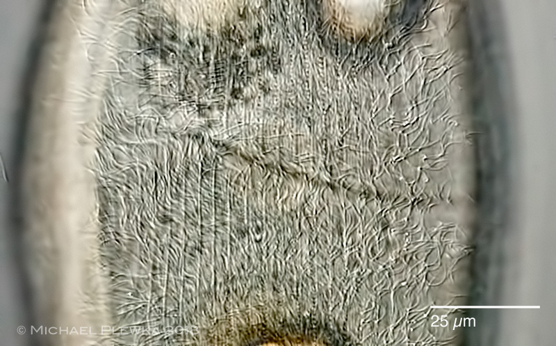
Turaniella vitrea; apikal cortex of a compressed specimen.
Turaniella vitrea; cilia in the area of the dividing fissure.
Turaniella vitrea; 3 aspects of thge macronucleus. Upper images: stained with methylene green; the dye affects the cell membrane / cortex so that the cytoplasm leaks (arrow). Lower image: stained with carmine- acetic acid.
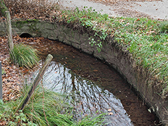
Location: Hattingen Wodantal, Heierbergsbach, puddle (click to enlarge)